The ALK gene encodes a membrane tyrosine kinase receptor and in humans it is located on the chromosome 2p23. The protein is composed of 1620 aminoacids and exhibits a tissue-specific expression, particularly in the brain, intestines and testis, but not in lymphocytes. Abnormlities in the ALK gene are, after the rearrangement of T-cell receptor, the most often found aberrations in T-lymphomas and are associated with nodal anaplastic large-cell lymphoma (ALCL). In 40 % ALCL there occurs translocation t(2;5)(p23;q35) resulting in the fusion of ALK with a nuclear phosphoprotein ALK (NPM). This fusion occurs in 70 - 80 % ALK positive ALCL. Other fusion partners of ALK are TPM3 located at 1q21 (10 - 20 % ALK positive ALCL), TGF located at 3q21 (2 - 5 %), ATIC located at 2q35 (2 - 5 %), CLTC located at 17q23 (1 - 5 %) and MSN located at Xq11-12 (< 1 %). All these rearrangements cause the overexpression of the ALK protein which may be detected by RT-PCR and also by immunohistochemistry, because normally there is no ALK expression in lymphatic cells. ALK expression can then be nuclear and/or cytoplasmic, depending on the fusion partner. However, the most sensitive detection method of ALK rearrangement represents FISH analysis with the break-apart designed probes.
Although the abnormal expression of the protein and ALK gene aberrations occur mostly in ALCL, they were described also in a normal nonhematopoietic tissue, in some rare B-lymphomas and in certain tumors of soft tissues, such as inflammatory myofibroblastic tumor.
Examination
Our laboratory has at its disposal ALK (Anaplastic Lymphoma Kinase) Dual Color, Break Apart Rearrangement probe from the Vysis/Abbott company which is designed as two different labeled probes hybridizing at a point of break of the ALK gene. Two fusion yellow signals per nucleus represent a normal finding. When the ALK gene breaks and subsequent translocation occurs we observe one orange, one green and one yellow signal.
FISH
When FISH, we detect the break of the gene ALK using ALK Dual Color, Break Apart Rearrangement Probe, Vysis/Abbot (this probe do not identify specific translocation partner). In a sample positive for rearrangement of the gene ALK we observe one yellow, one red and one green signal (fig. 1). In a negative sample we observe two yellow signals (fig. 2). (dual color filter)
-
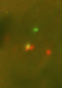 Fig.1
Fig.1Nucleus positive for the ALK gene breakage.
-
 Fig.2
Fig.2Nucleus negative for the ALK gene breakage.
References
- Kutok JL, Aster JC.:Molecular biology of anaplastic lymphoma kinase-positive anaplastic large-cell lymphoma. J Clin Oncol. 2002 Sep 1;20(17):3691-702. Review.
- Morris SW, Kirstein MN, Valentine MB, Dittmer KG, Shapiro DN, Saltman DL, Look AT. Fusion of a kinase gene, ALK, to a nucleolar protein gene, NPM, in non-Hodgkin's lymphoma.Science. 1994 Mar 4;263(5151):1281-4. Erratum in: Science. 1995 Jan 20;267(5196):316-7.