Gliomas are the most common primary tumors of the central nervous system. In terms of morphological appearance, biological behavior, genetic changes and clinical outcome they represent a very heterogeneous group. The most occurring are astrocytomas of which the most prevalent is highly malignant glioblastoma multiforme. A group of oligodendroglial tumors (OT) represents 5 - 18 % of all gliomas and includes classic oligodendrogliomas and mixed oligoastrocytomas. WHO distinguishes grade II and grade III (anaplastic) lesion (fig. 1). OT are, compared to other gliomas, predominantly typical for less tumor aggressiveness, slower progression and specific clinical features such as greater sensitivity to chemotherapy PVC (procarbazine, CCNU, vincristine) and longer survival of patients. However, pathogenesis is still insufficiently studied. Genetic characteristic of these tumors is combined loss of chromosome arms 1p and 19q as it was proven by FISH and LOS analysis in 60 to 90 % OT. Loss of 1p/19q is associated with longer survival, better response to radiotherapy and less toxic chemotherapy (temozolomide). Some authors put in relation the loss of 1p/19q only with better response of tumor to treatment regardless a tumor morphology.
Frequent loss of 1p/19q in OT suggests that these chromosomal arms may contain a tumor-suppresor gen (TSG) whose inactivation is included in the genesis of OT. However, concrete TSG has not yet been identified. Among candidate genes belong e.g. CAMTA1 (calmodulin-binding transcriptor activator 1) at 1p36, CDKN2C (cyklin-dependent dinase inhibitor 2C) localized at 1p32, p190RhoGAP gen (Rho kinase regulator) at 19q13.3 či EMP3 (myelin-related epithelial membrane protein) at19q13.3.
Besides the fact that testing of the loss 1p/19q in all oligodendrogliomas and tumors with oligodendroglial features it provides also prognostic and therapeutic information. Identification of 1p/19q codeletion can be useful for distinguishing OT from rare tumors such as dysembryoplastic neuroepithelial tumor, central neurocytoma, extraventricular neurocytoma or clear cell ependymoma. Deletion of 1p/19q has not been detected in these entities except extraventricular ependymoma which, according to some studies, suggests its possible histogenetic association with OT.
Examination
Determination of the loss 1p/19q is examined mainly by LOH analysis and fluorescent in situ hybridization (FISH). For this purpose, in our laboratory we use 2 different locus specific FISH probes 1p36/1q25 and 19q13/19p13. The result of this hybridization in negative sample are two green and two red signals per nucleus (fig. 2). In the case of loss of 1p or 19q we observe two green and one red signal (fig. 3).
-
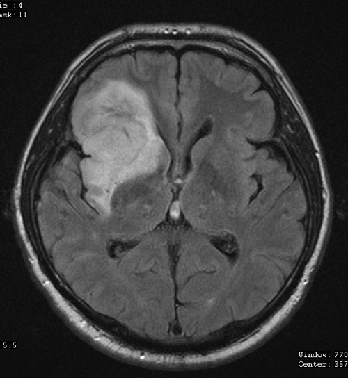 Fig.1
Fig.1Recording of magnetic resonance; anaplastic oligodendroglioma (courtesy of MUDr. Jan Mraček).
-
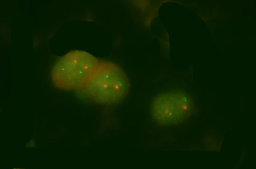 Fig.2
Fig.2Detection of the loss 1p. Example of the sample with a normal finding.
-
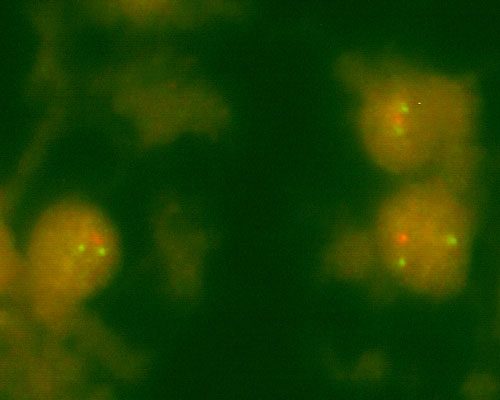 Fig.3
Fig.3Example of the loss 19q. Example of the sample with the loss of 19q.
References
- Cairncross JG, Ueki K, Zlatescu MC, Lisle DK, Finkelstein DM, Hammond RR, Silver JS, Stark PC, Macdonald DR, Ino Y, Ramsay DA, Louis DN. Specific genetic predictors of chemotherapeutic response and survival in patients with anaplastic oligodendrogliomas.J Natl Cancer Inst. 1998 Oct 7;90(19):1473-9.
- Smith JS, Alderete B, Minn Y, Borell TJ, Perry A, Mohapatra G, Hosek SM, Kimmel D, O'Fallon J, Yates A, Feuerstein BG, Burger PC, Scheithauer BW, Jenkins RB.Localization of common deletion regions on 1p and 19q in human gliomas and their association with histological subtype.Oncogene. 1999 Jul 15;18(28):4144-52.