Translocation t(8;14) (q24;q32) resulting in the fusion of the gene C-MYC and the gene for a heavy chain of immunoglobuline (IgH) presents the most common aberration that occurs in Burkitt's lymphoma (BL). In the remaining cases of BL we find especially t(8;22) (q24;q11) resulting in the fusion of the gene C-MYC and the gene for a light chain kappa or t(2;8) (p11;q24) that leads to the fusion of the gene C-MYC and the gene for a light chain lambda. C-MYC is a transcription factor that plays an important role in the cell cycle regulation, apoptosis and cell transformation. Therefore, deregulation of its expression is a significant factor of the neoplastic process.
The exact location of translocation differs in endemic and sporadic BL. Translocation t(8;14) in endemic BL occurs at the 5’ end at a distance up to 300 kb before the C-MYC coding region. In sporadic BL, translocation characteristically involves the point of break inside the gene C-MYC. The same translocations may also occur in Burkitt like lymphomas and in the small percentage in diffuse large B-cell lymphomas.
Simultaneously detected translocations C-MYC and t(14;18) are possible to find in the part of atypical BL arising from the follicular lymphoma. These lymphomas are then characterized by the aggressive disease course.
Currently, the most used method for detecting translocations involving C-MYC is fluorescent in-situ hybridization (FISH). The use of PCR and Southern blot is in the minority.
Examination
In our laboratory, we have 2 different FISH probes from the Vysis/Abbott company. The result of hybridization in a normal nucleus using LSI IGH/MYC, CEP 8 Tri-color, Dual Fusion Translocation probe are two aqua, two green and two orange signals. In the case of t(8;14) we can see two aqua, singly green and orange and one fusion signal per nucleus. Normal finding in the case of LSI MYC Dual Color, Break Apart Rearrangement probe are two fusion signals and in the case of translocation t(8;22)(q24;q11) or t(2;8)(p11;q24 we observe one orange, one green and one fusion signal per nucleus.
FISH
When FISH, we detect the break of the gene MYC using MYC Dual Color, Break Apart Rearrangement Probe, Vysis/Abbot (this probe do not identify specific translocation partner). In a sample positive for rearrangement of the gene MYC we observe one yellow, one red and one green signal (fig. 1). In negative sample we observe two yellow signals (fig. 2). (dual color filter)
-
 Fig.1
Fig.1Nucleus positive for the MYC gene breakage.
-
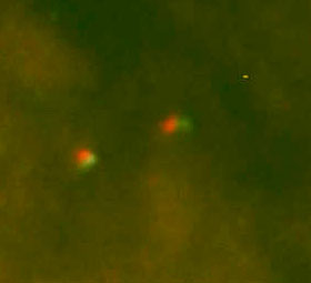 Fig.2
Fig.2Nucleus negative for the MYC gene breakage.
References
- Neri A, Barriga F, Knowles DM, Magrath IT, Dalla-Favera R.Different regions of the immunoglobulin heavy-chain locus are involved in chromosomal translocations in distinct pathogenetic forms of Burkitt lymphoma.Proc Natl Acad Sci U S A. 1988 Apr;85(8):2748-52.
- Busch K, Borkhardt A, Wossmann W, Reiter A, Harbott J.Combined polymerase chain reaction methods to detect c-myc/IgH rearrangement in childhood Burkitt's lymphoma for minimal residual disease analysis.Haematologica. 2004 Jul;89(7):818-25.